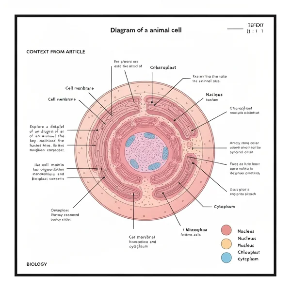
An animal cell diagram illustrates the internal structure and various organelles, such as the cell membrane, nucleus, mitochondria, and cytoplasm, which perform specific functions vital for the cell’s survival and operation. It helps in visualizing how these components interact and contribute to cellular processes like energy production and genetic information storage.
📌 Key Takeaways
Main purpose of this diagram: To visualize the intricate internal structure and organelles of an animal cell, aiding in the understanding of cellular biology.
Most important component to identify: The nucleus, as it houses the genetic material (DNA) and controls cell growth, metabolism, and reproduction.
Safety or critical consideration: Accurately distinguishing between animal and plant cell diagrams; animal cells lack a cell wall and chloroplasts.
Practical application tip: Use the diagram to label structures and describe their functions, reinforcing learning for biology exams or research.
When to use this diagram: When studying eukaryotic cell structure, comparing cell types, or explaining fundamental biological processes at the cellular level.
Diagram of an Animal Cell – Complete Guide
Whether you’re a student, a curious learner, or someone needing a quick reference, having a clear and accurate diagram of a animal cell is fundamental to understanding the basic unit of life. This guide is designed to clarify the intricate world within every animal, helping you decipher its vital components and their functions. From the protective outer layer to the energy-generating powerhouses, we’ll break down everything you need to know. By the end of this article, you will not only be able to identify each organelle but also understand its crucial role, turning complex biological structures into easily digestible knowledge.
Decoding the Animal Cell Diagram: A Visual Breakdown
An animal cell diagram serves as your essential map to the microscopic universe that constitutes all animal life. Typically depicted as a roughly spherical or irregular shape, it beautifully illustrates the various organelles suspended within the cytoplasm. The outermost boundary, the cell membrane, is often shown as a thin, flexible line, emphasizing its selective permeability. Dominating the center is usually the large, spherical nucleus, the cell’s control center, often with a darker nucleolus inside. Scattered throughout the cytoplasm, you’ll find numerous other structures, each with a distinct shape.
The bean-shaped mitochondria, often with internal folds (cristae), are readily identifiable as the powerhouses. Tiny dots represent ribosomes, sometimes free-floating or attached to the endoplasmic reticulum. The endoplasmic reticulum itself appears as a network of interconnected membranes, either rough (with ribosomes) or smooth. The Golgi apparatus, a stack of flattened sacs, is usually located near the nucleus. Smaller, often circular structures like lysosomes and small vacuoles (which are typically small and numerous in animal cells, unlike the large central vacuole in plant cells) complete the picture. Different diagrams may use color-coding to distinguish organelles, enhancing clarity and ease of identification for students and professionals alike.
💡 Key Information
Unlike plant cells, an animal cell diagram will not feature a rigid cell wall or chloroplasts. This is a primary distinguishing characteristic.
Diagram of an Animal Cell – Complete Guide
(Imagine a clear, well-labeled diagram of a typical animal cell here, showcasing its key organelles.)
Step-by-Step Guide: Interpreting Your Animal Cell Diagram
Related: diagram of a animal cell
Understanding an animal cell diagram is more than just memorizing labels; it’s about comprehending the function and interrelationship of each part. Follow these steps to effectively read and interpret any animal cell diagram, transforming it into a powerful learning tool.
1. Identify the Cell Membrane: The Outer Boundary
Start by locating the outermost layer. This is the cell membrane, also known as the plasma membrane. It’s typically shown as a thin, flexible line. Its primary role is to regulate what enters and exits the cell, acting as a selective barrier. Pay attention to any embedded proteins or surface markers that might be depicted, as these play roles in communication and transport.
2. Locate the Nucleus: The Cell’s Control Center
Next, find the largest and most prominent organelle, the nucleus. It’s usually centrally located and appears spherical or oval. Inside, you’ll often see a denser region called the nucleolus, involved in ribosome synthesis. The nucleus houses the cell’s genetic material (DNA), depicted as chromatin threads, and controls all cellular activities. The nuclear envelope, a double membrane with pores, encloses it.
3. Explore the Cytoplasm and Organelles: The Inner Environment
The jelly-like substance filling the cell and surrounding the organelles is the cytoplasm. Within it, you’ll find various specialized structures collectively known as organelles. These are the “little organs” that carry out specific functions necessary for the cell’s survival. Understand that the cytoplasm is not empty; it’s a bustling environment where many metabolic reactions occur.
4. Identify Mitochondria: The Energy Generators
Look for the oval or bean-shaped organelles with distinctive inner folds called cristae. These are the mitochondria, often referred to as the “powerhouses of the cell.” They are responsible for cellular respiration, generating ATP (adenosine triphosphate) – the cell’s main energy currency. Their unique internal structure is key to their function.
5. Spot Ribosomes, ER, and Golgi: Protein Synthesis & Modification
✓Ribosomes: Tiny dots, either free in the cytoplasm or attached to the Rough Endoplasmic Reticulum (RER). They are the sites of protein synthesis.
✓Endoplasmic Reticulum (ER): A network of interconnected sacs and tubules. The RER (with ribosomes) processes proteins, while the Smooth ER (SER, without ribosomes) is involved in lipid synthesis and detoxification.
✓Golgi Apparatus (or Golgi Complex/Body): Appears as a stack of flattened sacs (cisternae). It modifies, sorts, and packages proteins and lipids for secretion or delivery to other organelles.
6. Identify Other Key Organelles: Lysosomes & Vacuoles
Look for other smaller, membrane-bound sacs. Lysosomes are often small and spherical, containing digestive enzymes to break down waste materials and cellular debris. Vacuoles in animal cells are typically small and numerous, serving various storage and transport functions, distinct from the large, central vacuole found in plant cells. Centrioles, usually found near the nucleus, are involved in cell division.
Common Issues & Troubleshooting When Reading Diagrams
Related: diagram of a animal cell
Even with a clear diagram of a animal cell, certain difficulties can arise. Knowing these common pitfalls can help you troubleshoot your understanding and avoid misconceptions.
✓Confusing Animal vs. Plant Cells: The most frequent error is expecting features like a cell wall or chloroplasts in an animal cell. Remember, animal cells lack these structures, which are defining characteristics of plant cells. Always verify the cell type before proceeding.
✓Misidentifying Organelles: Some organelles, especially the ER and Golgi, can look similar at first glance. Pay close attention to their unique shapes (network vs. stack of sacs) and surface features (ribosomes on RER). Reviewing their specific functions helps solidify identification.
✓Understanding 2D Representation: Cell diagrams are often 2D representations of complex 3D structures. Remember that organelles exist in three dimensions within the cell. This can sometimes make interpreting their exact shape or spatial relationship challenging.
✓Overwhelmed by Detail: Highly detailed diagrams can be daunting. Start by identifying the major organelles first (cell membrane, nucleus, mitochondria), then gradually add the smaller structures. Don’t try to absorb everything at once.
⚠️ Warning
Always cross-reference with multiple reliable sources if a diagram seems unclear or contradictory. Discrepancies can sometimes occur in simplified or older diagrams.
Tips & Best Practices for Mastering Animal Cell Diagrams
To truly master the diagram of a animal cell and the biology it represents, active learning and strategic study methods are key. Incorporate these tips into your routine for better retention and deeper understanding.
✓Active Labeling and Drawing: Don’t just look at diagrams; interact with them. Print out unlabeled diagrams and try to label them yourself. Even better, draw your own animal cell diagram from memory, adding each organelle as you recall its function. This actively engages your brain in retrieval practice.
✓Relate Structure to Function: Always ask “why does it look this way?” For example, the extensive folding of mitochondria’s inner membrane (cristae) increases surface area for energy production. Understanding these structure-function relationships makes memorization easier and more meaningful.
✓Use Multiple Perspectives: Look at diagrams from different textbooks or online resources. While the core components remain the same, variations in rendering can highlight different features and provide a more comprehensive mental model of the cell.
✓Create Mnemonics and Analogies: Develop memory aids for the organelles and their functions. For instance, the nucleus is the “brain,” mitochondria are the “power plants,” and ribosomes are “protein factories.” Analogies help connect new information to familiar concepts.
✓Review Regularly: Biology concepts, especially those involving detailed structures, benefit from consistent review. Short, frequent study sessions are more effective than long, infrequent ones.
✅ Pro Tip
Utilize interactive 3D cell models online or in apps. These can significantly enhance your understanding of the spatial relationships between organelles, a common challenge with static 2D diagrams.
By thoroughly engaging with a diagram of a animal cell and applying these strategies, you’ll not only commit its parts to memory but also gain a deeper, functional understanding of this fundamental biological unit. This comprehensive approach will serve you well in any scientific endeavor, from introductory biology to advanced cellular research.
Frequently Asked Questions
What is an animal cell diagram?
An animal cell diagram is a visual representation showcasing the various organelles and structures within an animal cell. It typically labels components like the cell membrane, nucleus, mitochondria, and cytoplasm, illustrating their relative positions and shapes. These diagrams are crucial educational tools for understanding basic cellular biology and the functions each part performs.
How do you read an animal cell diagram?
To read an animal cell diagram, first identify the main cell boundary, the cell membrane. Then, locate the prominent nucleus, usually central. Proceed to identify other organelles like the mitochondria, endoplasmic reticulum, and ribosomes in the cytoplasm. Pay attention to labels and arrows that indicate specific structures and their roles, connecting form to function.
What are the parts of an animal cell?
An animal cell comprises several vital parts. Key components include the flexible cell membrane, the genetic control center known as the nucleus, and the jelly-like cytoplasm filling the cell. Other crucial organelles are mitochondria (for energy), ribosomes (for protein synthesis), endoplasmic reticulum, Golgi apparatus, and lysosomes, all working synergistically.
Why is the nucleus important?
The nucleus is critically important because it houses the cell’s genetic material, DNA, organized into chromosomes. It controls all cellular activities, including growth, metabolism, protein synthesis, and reproduction, by regulating gene expression. Essentially, the nucleus acts as the command center, ensuring the cell performs its correct functions and maintains its integrity.
What is the difference between animal and plant cells?
The main differences between animal and plant cells lie in a few key structures. Animal cells lack a cell wall, chloroplasts, and a large central vacuole, which are characteristic of plant cells. Plant cells have a rigid cell wall for support and chloroplasts for photosynthesis. Animal cells have centrioles, which are typically absent in plant cells.
How do I use an animal cell diagram?
Use an animal cell diagram to learn and reinforce knowledge of cellular anatomy and physiology. Begin by identifying and labeling each organelle. Then, research and describe the specific function of each component, such as the cell membrane’s role in transport or mitochondria’s role in energy production. This practice helps deepen your understanding of cell biology.
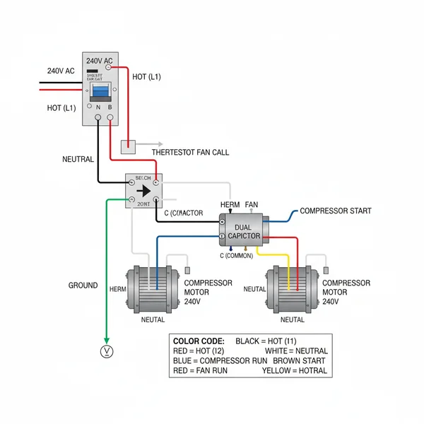
A 3 wire AC dual capacitor wiring diagram illustrates how to connect a dual-run capacitor to an HVAC unit’s fan and compressor motors. It details connections for the common terminal, herm, and fan terminals, showing proper routing for the hot wire and neutral wire. This ensures safe and efficient startup and continuous operation of both…
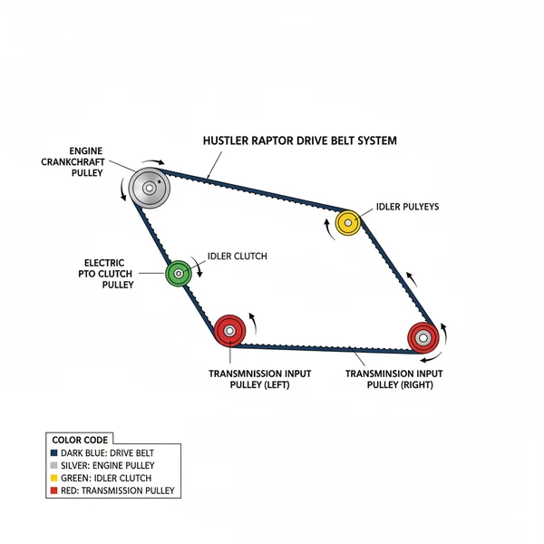
The Hustler Raptor drive belt diagram visually illustrates the component layout of the mower’s drive system, showing belt routing around pulleys and tensioners. It’s crucial for understanding the system’s configuration, aiding in correct belt replacement, tension adjustment, and overall maintenance to ensure optimal mower performance. 📌 Key Takeaways Main purpose of this diagram is to…
The 5.7 Chevy 350 belt diagram with AC illustrates the serpentine belt’s path around various engine components like the crankshaft, alternator, power steering pump, and AC compressor. It’s crucial for understanding the correct configuration of your vehicle’s accessory drive system and ensuring proper function of all connected accessories. 📌 Key Takeaways Main purpose of this…
The Mass Air Flow (MAF) sensor is typically located in the engine’s intake tract, between the air filter box and the throttle body. It measures the amount of air entering the engine, sending this crucial data to the ECU for precise fuel mixture calculations. Identifying its location is vital for maintenance and troubleshooting. 📌 Key…
The Chevy 4.3 V6 engine diagram visually details all major engine components, their arrangement, and connections. It is invaluable for understanding the engine’s layout, performing maintenance, locating parts for replacement, and troubleshooting issues. This resource helps mechanics and DIYers efficiently identify problem areas and plan repairs. 📌 Key Takeaways To visualize and understand the complex…
This diagram illustrates the ideal configuration for grab bar placement in a walk-in shower, ensuring maximum safety and accessibility. It details the precise height, angle, and orientation of each essential component to create a secure structure, preventing falls and supporting user independence within the shower system. 📌 Key Takeaways Main purpose of this diagram: To…